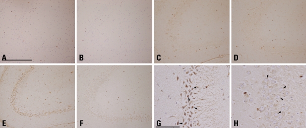
Fig. 4.
Immunohistochemistry of cathepsin E in rat hippocampus. After i.p. administration of normal saline (A and B) or KA (12 mg/kg in C and D, 25 mg/kg in D, E, G and H), rats were fed a normal diet (A, C, E and G) or the ketogenic diet (B, D, F and H). The CA3 region of the rat hippocampus of the NS-ND (A) and the NS-KD (B) equally showed very few cathepsin-E positive neurons. In the lower-dose KA-ND rat (C), a small number of neurons expressed cathepsin E, while those of the lower-dose KA-KD rat (D) distributed more sparsely. In the CA3 region of the higher-dose KA-ND rat (E), widespread expression of cathepsin E by neurons were observed, in contrast to a marked decrease in the cathepsin E immunoreactivity in the same area of the higher-dose KA-KD rat (F). Details of higher-dose KA-ND neurons exhibit pyknotic changes (G, arrowheads), whereas neurons of the higher-dose KA-KD rat had round, euchromatic nuclei (H, arrowheads). A, B, G, and H were counterstained (scale bar = 500 µm in A, 100 µm in G; A through F are in a same degree of magnification, so are G and H). KA, kainic acid; NS-ND, NS-ND, normal saline, normal diet; NS-KD, normal saline, ketogenic diet; KA-KD, kainic acid, ketogenic diet; CA3, cornu ammonis.

