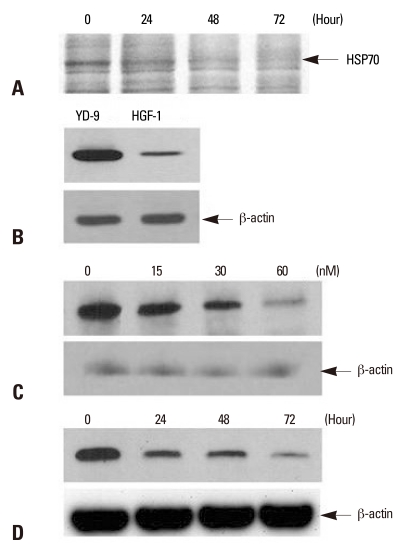
Fig. 2.
Detection of HSP70 and the effect of PEA on HSP70 expression in YD-9 cells. (A) After incubation with15 nM PEA for indicated periods, identification of differentially expressed proteins using LC-MS/MS indicated HSP70. (B) Western blot for HSP70 from YD-9 and HGF-1 cells was performed. The expression level of HSP70 in YD-9 cells was higher than HGF-1 cells. (C) After incubation with indicated doses of PEA for 24 hours, Western blot analysis showed a decrease of HSP70 expression. (D) After incubation with 15 nM of PEA for indicated time periods, Western blot analysis showed a time-dependent continual decrease in HSP70 expression.

