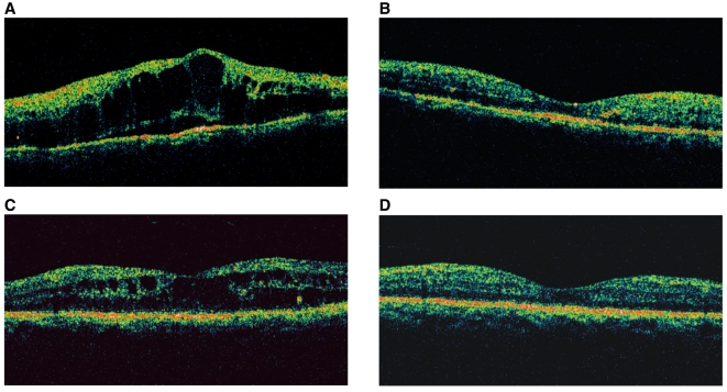
Fig. 3.
Changes in the OCT images of representative patient in the intravitreal and posterior subtenon injection groups. In an eye that underwent intravitreal injection, the marked macular edema (A) decreased substantially and the eye showed virtually normal macular configuration at 3 months (B) after injection. In an eye that underwent posterior subtenon injection, the macular edema (C) also decreased substantially with time, and the eye showed virtually normal macular configuration at 3 months (D) after injection.

