
Figure 5.
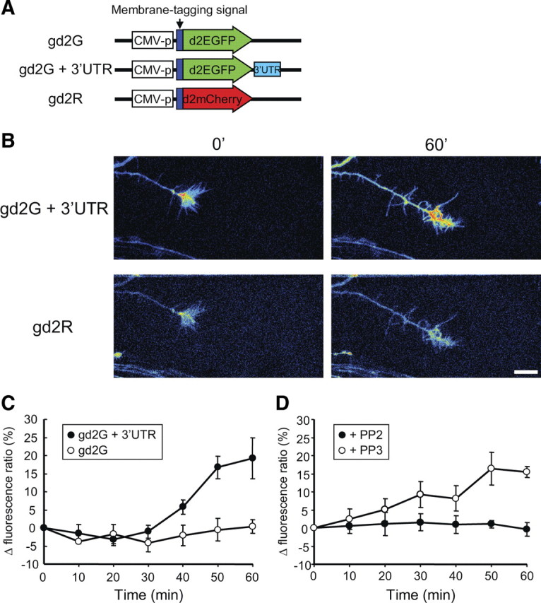
The Src family kinase inhibitor PP2 suppressed BDNF-induced local translation in growth cones using a GFP reporter with the 3′ UTR of β-actin. Neurons were transfected with designated reporters and cultured as neuron balls extending axons >1 mm in length (see Materials and Methods) (supplemental Fig. S6, available at www.jneurosci.org as supplemental material). A, Vector structures of fluorescent reporters. Both GFP and mCherry reporters were fused with a membrane-targeting sequence (dual palmitoylation from GAP43) and a sequence for shorter half-life (d2EGFP and d2mCherry). The GFP reporter (gd2G) contains the 3′ UTR sequence of rat β-actin, but the mCherry reporter (gd2R) does not contain this sequence. CMV, Cytomegalovirus. B, Representative images of growth cones of cortical neurons in neuron ball cultures cotransfected with gd2G + 3′ UTR and gd2R. Pseudocolored images show that the absence of fluorescent signal is indicated by black, with increasing fluorescence indicated by transitions to blue, green, yellow, and red. Note that BDNF stimulation increased the fluorescence of gd2G + 3′ UTR, but not gd2R, after 60 min. Scale bar, 10 μm. C, Effect of the β-actin 3′ UTR on expression of the GFP reporter in growth cones. The GFP reporter (gd2G or gd2G + 3′ UTR) was cotransfected with gd2R into cortical neurons. Neurons were stimulated with BDNF, and fluorescence ratios of the GFP reporters to gd2R were calculated at indicated times and then normalized to the initial fluorescence ratio. The fluorescence ratio of gd2G+3′ UTR to gd2R increased from 30 min after BDNF stimulation, but the fluorescence ratio of gd2G did not. D, The SFK inhibitor PP2, but not its inactive derivative PP3, inhibited the increase of the fluorescence ratio of gd2G + 3′ UTR to gd2R after BDNF stimulation.