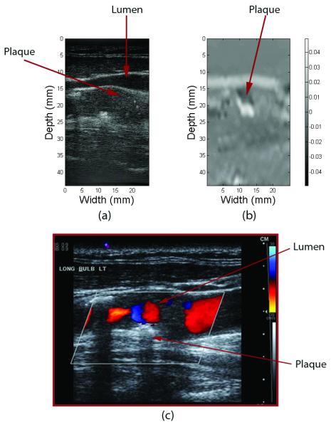
Figure 5.

(a) B-mode gray scale ultrasound image, (b) 2-D elastogram, (c) B-mode with colorflow of an in-vivo carotid artery with plaque visible near the wall of the vessel close to the bifurcation. Note also the presence of turbulent flow around this region. Elastographically strain here appears as a more friable or more likely to fracture region and is depicted as the brighter region.
