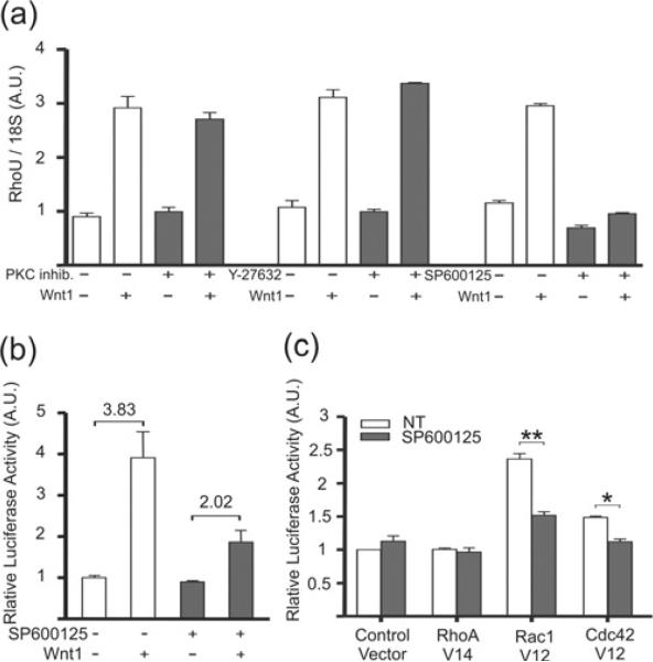
Figure 5. Wnt-1-mediated RhoU activation follows a non-canonical pathway involving JNK.
(a) qRT-PCR analysis. Wild-type MEFs were stimulated or not with Wnt-1 as described for Figure 3 and treated with specific inhibitors for PKC (myristoylated peptide inhibitor, 100 μM), ROCK (Y-27632, 10 μM) and JNK (SP600125, 50 μM) for 24 h. RhoU expression levels were measured by qRT-PCR and normalized to 18S RNA. Values are means ± S.E.M. of two independent experiments performed in duplicate. (b and c) Transient transfection assays. Wild-type MEFs were transfected with the promoter fragment −770 alone (b) or in combination with plasmids encoding the constitutively active GTPases RhoA (RhoA V14), Rac1 (Rac1 V12), Cdc42 (Cdc42 V12) or a control empty vector (c). At 24 h later, cells were stimulated or not with Wnt-1 as described for Figure 3 and treated with 50 μM SP600125 (b) or simply treated or not with 50 μM SP600125 (c) and cultured for a further 24 h. Luciferase activity was quantified and normalized as described for Figure 2. A.U., arbitrary units.

