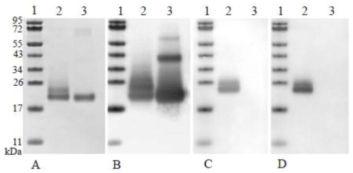
Figure 2.
Comparison of ISOkine Flt3 ligand and an E. coli produced Flt3 ligand from commercial source. Lane 1 contains a molecular weight marker, lane 2, purified ISOkine Flt3 ligand and in lane 3 E. coli produced Flt3 ligand from commercial source. Figure A shows Coomassie blue stained SDS-PAGE gel. Six hundred nanogram was loaded on the gel. Figure B–D show Western blot analysis using polyclonal anti-Flt3 ligand antibodies (B), anti-α-1,3-fucose specific antibody (C) and anti-β-1,2-xylose specific antibody (D). Two hundred and fifty nanogram of protein was loaded on the gel for the Western blot application.

