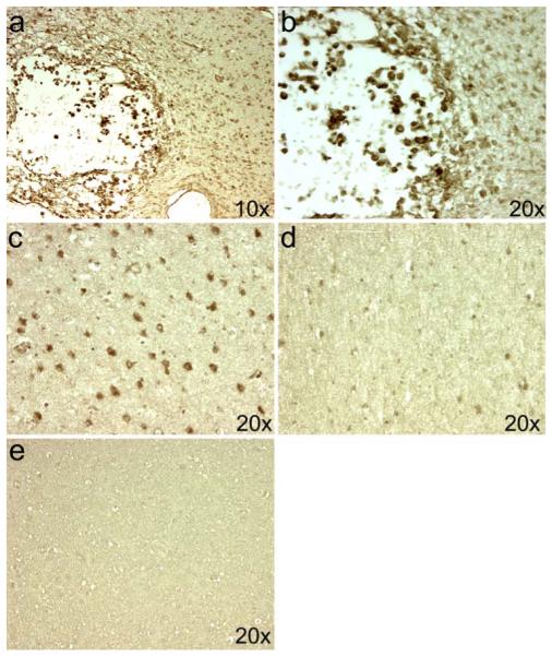
Fig. 1.

Localization of iNOS expression in PVL. The expression of iNOS is seen in macrophages of the focal necrosis of a subacute PVL lesion in a 39-PC week infant (a, b). In this same case, iNOS expression is seen in the diffusely gliotic white matter surrounding the focal necrosis in cells morphologically identified as reactive astrocytes (c). The number of iNOS-expressing cells is significantly reduced in a control case at 37 PC weeks (d). Binding of the iNOS antibody was blocked in the PVL case from above using an excess of the peptide from which the antibody was produced (e)
