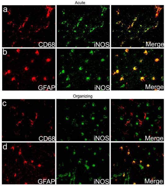
Fig. 3.

Cell-specific expression of iNOS in the diffuse component of PVL. The expression of iNOS is seen in CD68-positive microglia (a) as well as in GFAP-positive reactive astrocytes (b) in the diffuse component of an acute PVL case at 40 PC weeks. A PVL case of the subacute (organizing) phase at 39 PC weeks shows no iNOS expression in CD68-positive microglia (c), but continues to show strong iNOS expression in GFAP-positive reactive astrocytes (d)
