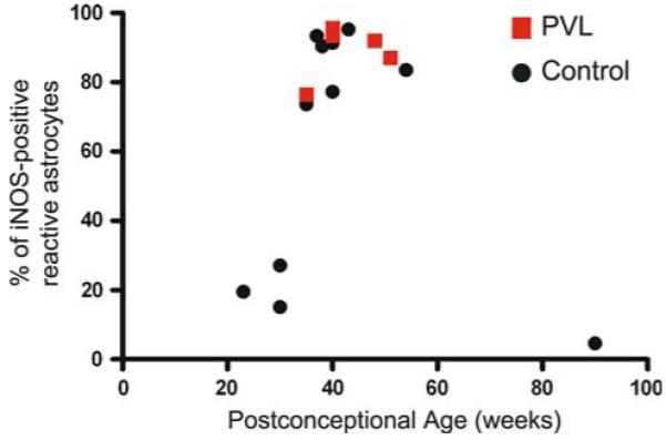
Fig. 4.

Astrocytic expression of iNOS in PVL and controls. The percentage of GFAP-positive reactive astrocytes that express iNOS was determined in the diffuse component of PVL and in the corresponding periventricular white matter of control cases. In the overlapping ages of 35–54 PC weeks, there is no significant difference in the percentage of GFAP-positive astrocytes that express iNOS, although the overall density of iNOS positive glia (Fig. 2) as well as GFAP positive astrocytes [16] in control tissue is significantly less than in PVL
