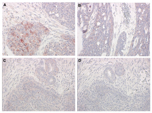
Figure 5.
Immunohistochemical staining for phosphorylated Akt. Sections of tumors were stained for phospho-Akt (A, B, C) or control rabbit IgG (D) and counterstained with hematoxylin. Genotypes of mice were Wnt-1 TG+, Pten+/- (A, B); Wnt-1 TG+, Pten+/+ (C, D). (A) and (B) were from tumors with or without LOH, respectively. A 40x objective was used.

