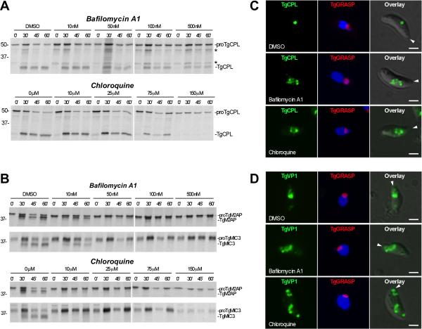
Fig. 8. In vivo maturation of proTgCPL and proTgM2AP are differentially sensitive to pH antagonists.
A. Representative autoradiographs showing inhibition of processing in parasite treated with pH-altering substances. RH parasites were pretreated for 15 min with the indicated concentrations of bafilomycin A1 or chloroquine before pulse labeling with [35S] Met/Cys for 10 min, chasing for the indicated times, and immunoprecipitation with Rαr100TgCPL. Asterisks indicate intermediate processed forms (48 kDa and 32 kDa) of proTgCPL.
B. Same as A except immunoprecipitations were performed with RαrTgM2AP and mAb T4.2F3 against TgMIC3. Molecular weight markers are expressed in kDa.
C. Bafilomycin A1 and chloroquine induced enlargement of the VAC. Representative immunofluorescence images showing TgCPL localization in RH parasites expressing TgGRASP-mRFP after treatment with pH antagonists. Parasites were treated with DMSO (upper panel); 500 nM bafilomycin A1 (middle panel); or 150 μM chloroquine (lower panel) for 15 min at RT, and then incubated for another 1h at 37°C.
D. Bafilomycin A1 and chloroquine induced swelling and disassembling of the LE. Representative immunofluorescence images showing TgVP1 localization in RH parasites expressing TgGRASP-mRFP after treatment with DMSO (upper panel); 500 nM bafilomycin A1 (middle panel); or 150 μM chloroquine (lower panel) as described above. Arrowheads indicate the apical end of the parasite.

