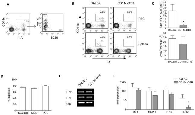
Figure 4. Dendritic cells are not required for IFN-I production induced by TMPD.
A) Flow cytometry of peritoneal DCs after TMPD treatment. Box indicate CD11c+ I-A+ DCs. B) Depletion in CD11c-DTR mice 2 days after diphtheria toxin (DT) injection. Box indicates CD11c+ I-A+ DCs. C) Quantification of peritoneal dendritic cells and Ly6Chi monocytes. D) Quantification of splenic DC depletion. MDCs were defined as CD11chi I-A+ CD11b+ cells and PDCs were defined as CD11c+ B220+ PDCA-1+. E) IFN-I expression (conventional PCR) and F) ISG expression (RT-PCR) in peritoneal exudates cells. Each bar represents the mean of 6 animals and error bars indicate s.d. * p < 0.05 (Student’s t-test).

