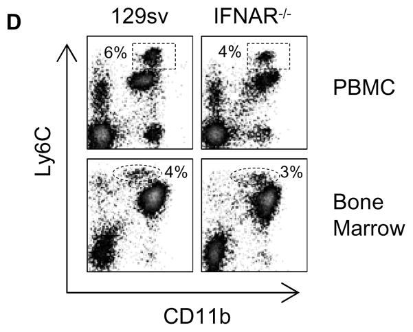
Figure 6. Ly6C expression on immature monocytes is not dependent on IFN-I signaling.
Flow cytometry analysis of monocyte subsets in the peripheral blood (upper) and bone marrow (lower) of TMPD-treated wild-type 129sv and IFN-I receptor-deficient mice. Boxes and ovals indicate peripheral blood Ly6Chi monocytes and bone marrow Ly6Chi monocyte precursors, respectively.

