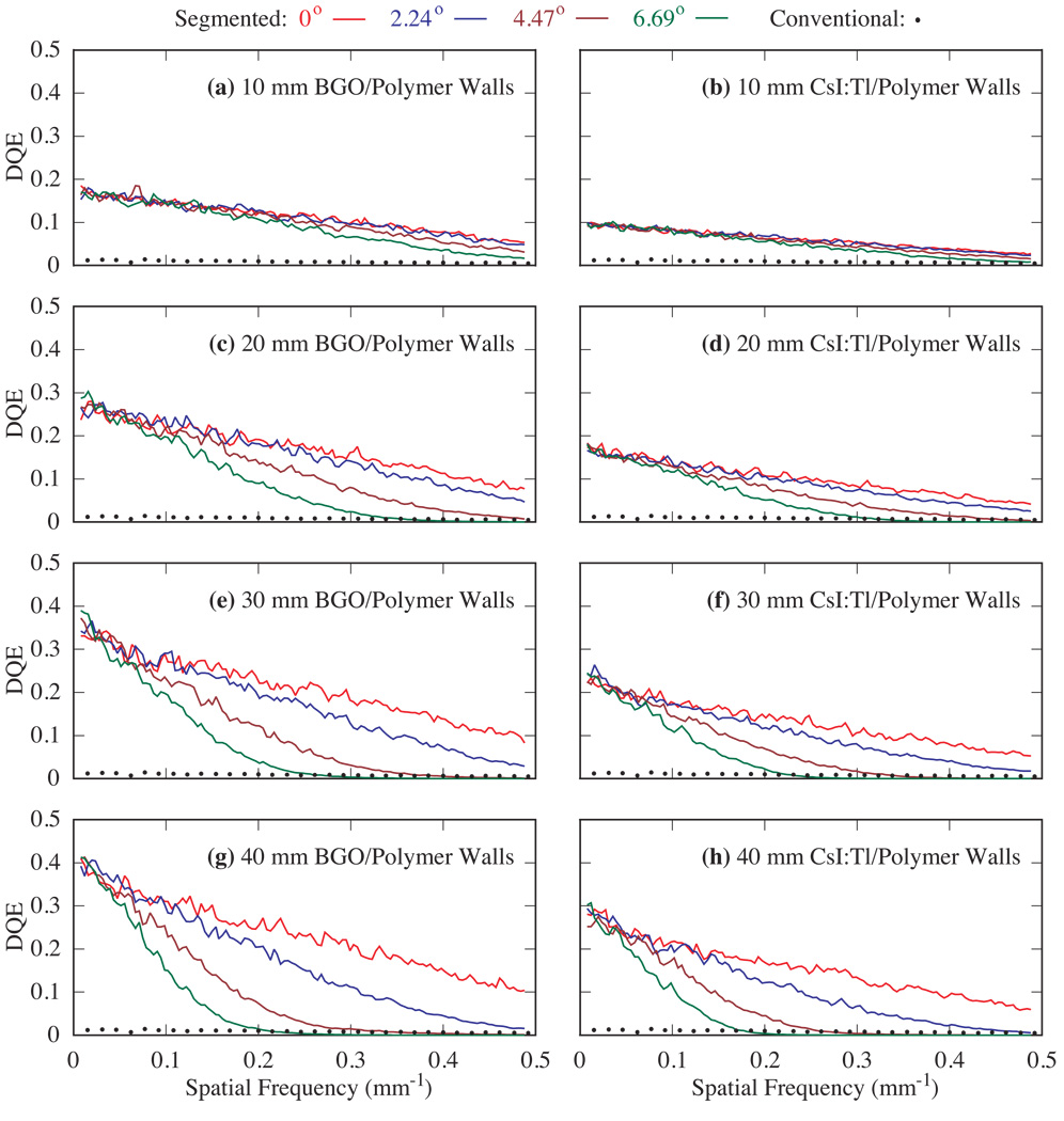
FIG. 5.
DQE for four incident beam angles (0°, 2.24°, 4.47°, and 6.69°) using 10 – 40 mm thick, segmented BGO and CsI:T1 detectors with polymer septal walls. In this figure and in figure 6, for each detector configuration, the progressive degradation in DQE shown by the various lines corresponds to increasing incident beam angle. Note that the results are compared to DQE measured from the conventional EPID.

