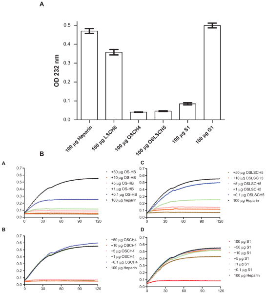
Figure 3.
A) Detection of contaminated heparin by enzymatic assay. Exactly 100 μg of heparin and the five contaminated heparin lots shown in Figure 1 were digested for 2 hr with 1 mU heparin lyase I in triplicate and the absorbance at 232 nm was recorded for 120 min. The maximum change in absorbance (OD) of 100 μg of digested samples is shown with standard deviation. B) Inhibition heparin digestion by chemically sulfated heparin byproduct and contaminated heparins. Heparin (100 μg) alone or heparin plus chemically oversulfated heparin byproduct (OS-HB) or other contaminated heparins (OSCH4, OSLSCH5, and S1) in duplicates were digested with heparin lyase I and the absorbance at 232 nm was recorded every min for a total 120 min. Each data point of 120 data points shown in each digestion curve was average values of two independent heparin lyase digestion reactions. A) 100 μg heparin plus 0, 0.1, 1, 5, 10, or 50 μg of oversulfated heparin byproduct; B) 100 μg heparin plus 0, 0.1, 1, 5, 10, or 50 μg of contaminated heparin OSCH4; C) 100 μg heparin plus 0, 0.1, 1, 5, 10, or 50 μg of contaminated heparin OSLSCH5; D) 100 μg heparin plus 0, 0.1, 1, 5, 10, or 50 μg of contaminated heparin S1.

