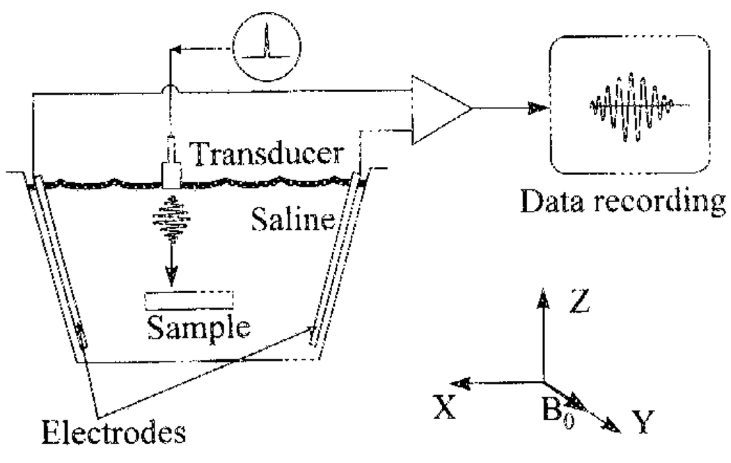
Fig. 2.
Diagram of the experiment to image with HEI a sample immersed in a saline chamber. The dimensions of the chamber are 27 cm (X dimension) × 17 cm (Y)× 22 cm (Z). The electrodes are two exposed copper wire segments. The piezoelectric ultrasound transducer has a center frequency of 1 MHz, and a 6-dB bandwidth of 0.6 MHz. An electrical pulser inputs single-phase electrical pulses of approximately 0.15-µs duration into the transducer, which in turn emits ultrasound pressure pulses into the chamber. The Hall voltage is amplified by 60-dB gain, filtered by a 0.1–3-MHz bandpass filter, and digitized at 5 megasample/s for data storage.

