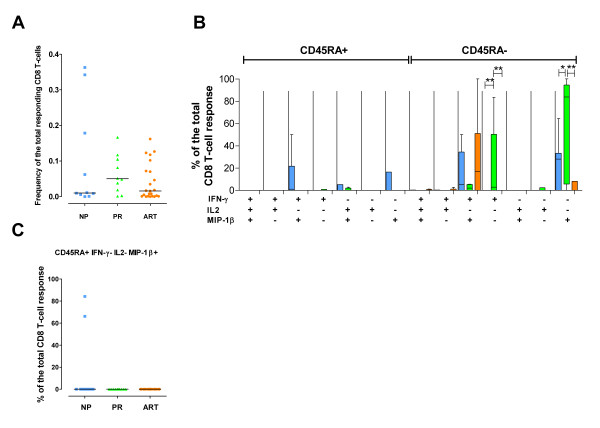
Figure 3.
HIV-1-Tat-specific CD8+ T-cell responses. HIV-1-Tat-specific CD8+ T-cell responses in 10 NP (blue), 10 PR (green) and 23 ART-treated patients (orange). Tat-specific responses were not analyzed in NP11. (A) Frequency of the total Tat-specific CD8+ T cells. (B) Quality of the Tat-specific response. The graph is divided into the CD45RA+ (left part) and the CD45RAneg (right part) CD8+ T-cell populations. All the possible combinations of the responses are shown on the x-axis for NP, PR and ART-treated individuals. Tukey boxes and whisker plots are shown. Significant differences are noted above the graph: (*) p < 0.05, (**) p < 0.01 and (***) p < 0.001. (C) Individual data point representation of the Tat-specific CD45RA+ IFN-γneg IL-2neg MIP-1β+ CD8+ T-cells. In all graphs, medians are represented by horizontal bars.

