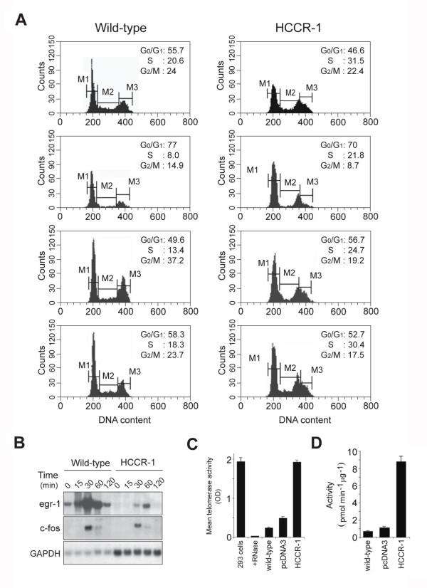
Figure 3.
Molecular alterations in HCCR-1-induced tumorigenesis. A. Cell-cycle profiles of wild-type NIH/3T3 cell and HCCR-1-transfected cells from a separate experiment. Exponentially growing cells were trypsinized and DNA content was determined by flow cytometry. To assess the serum-dependent cell cycle progression, cells were cultured in 0.5% BCS for 36 houes. After incubation, cells were released with 20% serum and harvested. B. Tumor suppressor egr-1 expressions. HCCR-1-transfected cells were serum starved with 0.5% BCS for 36 hours, and then stimulated with fresh 20% serum. Total RNA was isolated, transferred, and hybridized with 32P-labeled egr-1, c-fos, and GAPDH probe, respectively. C. Determination of telomerase activity. Human telomerase-positive embryonic kidney 293 cells, 293 cell extracts treated with RNase (+ RNase), wild-type NIH/3T3, cells transfected with vector alone and HCCR-1-transfected cells were analyzed by Telomerase PCR ELISA. Assays were performed according to the kit protocol with amounts of extracts equivalent to 1 ×10 3 cells. The telomerase activity in 293 cells, which served as a positive control, was abolished by pretreatment with RNase. Results are the average mean absorbance values from four separate experiments (means and 95% confidence intervals) (wild-type versus HCCR-1, P < .05). D. Determination of PKC activity. Wild-type NIH/3T3, cells transfected with vector alone and HCCR-1-transfected cell extracts were prepared and assayed to determine PKC activity. Each value is the means and 95% confidence intervals of three independent experiments (wild-type versus HCCR-1, P < 0.05).

