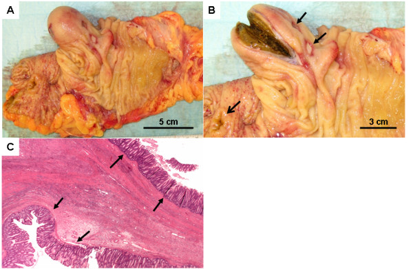
Figure 2.

Macroscopical and histopathological findings of the specimen. A) The right hemicolon opened with the cystic duplication. B) The stool-filled cystic lesion is cut open. Notice the mucosal ulcerations (closed arrows). The open arrow depicts the ileo-ceacal area. C) Histopathological demonstration of the duplication cyst.
