Fig. 1.
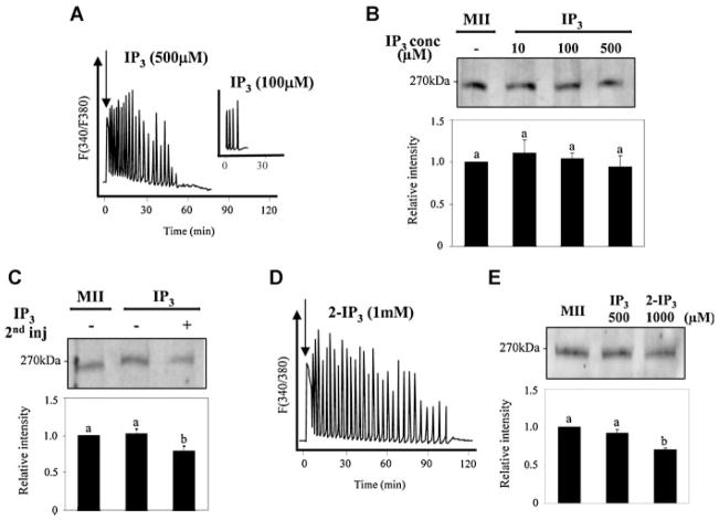
Bolus injection of IP3 that generates transient increases in [IP3]i induces [Ca2+]i oscillations that fail to promote substantive IP3R1 degradation. Injection of IP3 initiated dose-dependent [Ca2+]i responses in mouse eggs (A; 500 μM IP3; inset, 100 μM IP3). These IP3 concentrations (10, 100, and 500 μM; lanes 2, 3, and 4, respectively) did not induce noticeable receptor degradation in eggs collected 4 h post-injection (B). A double injection of 500 μM IP3, the second injection of which was delivered 2 h after the first injection, resulted in an ~20% loss of IP3R1 mass (C). Injection of a metabolically stable form of IP3, 2-IP3, at 1 mM initiated long-term oscillations (D) without inducing significant receptor degradation (E). The intensity of the immunoreactive bands in all parts in this figure (B, C, E; bar graphs) and elsewhere in the article was normalized to that of uninjected MII eggs, which was given the value of 1; bars with different superscripts represent values with significant differences (P < 0.05). Arrows in(A) and(D) denote time of injection. In(C), the minus sign under the IP3 treatment indicates single injection of IP3.

