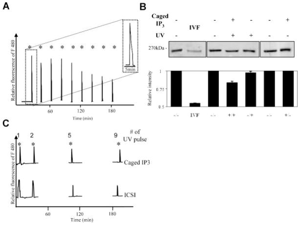
Fig. 2.
Discrete IP3 pulses from caged IP3 cause single [Ca2+]i rises and minor IP3R1 degradation. MII mouse eggs injected with caged IP3 were exposed to UV light for 0.005 sec at intervals of 20 min to cause pulsatile release of IP3. Each UV flash induced a [Ca2+]i rise that was detected in a [Ca2+]i monitoring system; 9 pulses were delivered per egg (A). The inset in (A) shows an enlarged version of the first rise so that the greater duration of the Ca2+ spike can be appreciated. The IP3R1 degradation induced by periodic release of IP3 was compared to that induced by IVF (B). Eggs were collected 4 h after the beginning of the uncaging protocol or 5 h after insemination. The intensity of IP3R1 degradation was quantified as described above. Simultaneous Ca2+ monitoring of [Ca2+]i rises #1, 5, and 9 induced by caged IP3 and by ICSI reveals that both procedures triggered[Ca2+]i rises of similar amplitude and duration (C). IP3R1-immunoreactive bands within each box (B) denote samples that were collected and processed for Western blotting at the same time. Given the number of samples needed for each experiment and the number eggs needed to perform Western blots, it was technically not possible to perform all sample collections in the same experiment.

