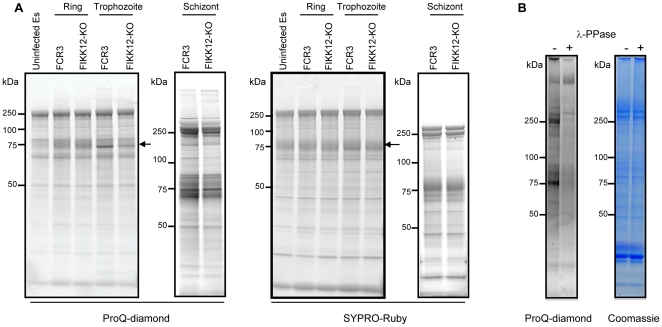
Figure 4. FIKK12-KO changes the phosphorylation pattern of a 80 kDa protein.
A. Phosphorylation pattern in the ghost fraction of FIKK12-KO parasite. Ghost fractions from uninfected erythrocytes and ring, trophozoite and schizont stages of FCR3 and FIKK12-KO IEs were subjected to SDS-PAGE. The gel was stained with ProQ-diamond (left panel) and then stained with SYPRO-Ruby (right panel). Arrow indicates a band of 80 kDa protein. B. Control of the specificity of ProQ-diamond. The ghost fraction from trophozoite stage of FCR3 was incubated with lambda protein phosphatase, subjected to SDS-PAGE, and stained with ProQ-diamond. The gel was then stained with Coomassie blue.

