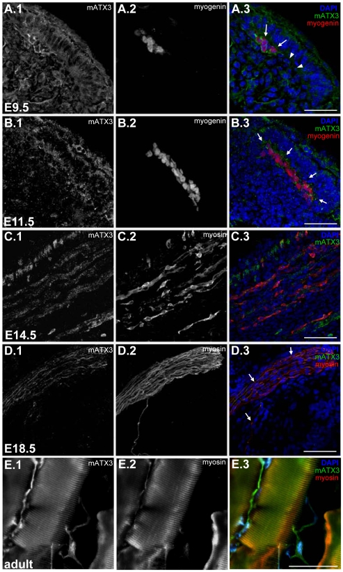
Figure 1. Mouse ataxin-3 (mATX3) is highly expressed in the early differentiated cells in the myotome,as well as in embryonic and adult muscle fibers.
Maximum projection of confocal optical z-series from sagittal 15 µm sections of E9.5 (A), E11.5 (B), E14.5 (C) and E18.5 (D) mouse embryos labeled for mATX3 and the myogenic differentiation markers myogenin (A, B) or myosin (C, D). (A) mATX3 (green, arrows) is highly upregulated in differentiated myogenin-positive cells (red) when comparing with younger myogenin-negative cells in the myotome (arrowheads). (B) strong staining for mATX3 (green, arrows) remains present in E11.5 elongated myogenin-positive myocytes (red). (C) E14.5 intercostal muscle showing a fiber-like labeling pattern of mATX3 (green) colocalising with myosin-positive fibers (red). (D) mATX3 (green, arrows) remains strongly expressed at late fetal stages in myosin-positive fibers (red). (E) mATX3 (green) is expressed in adult myosin fibers (red). (1) mATX3 staining, (2) myosin (MHC) or myogenin labeling, (3) triple overlay of mATX3, myogenic marker, and DAPI. Scale bars represent 80 µm.

