
Figure 7.
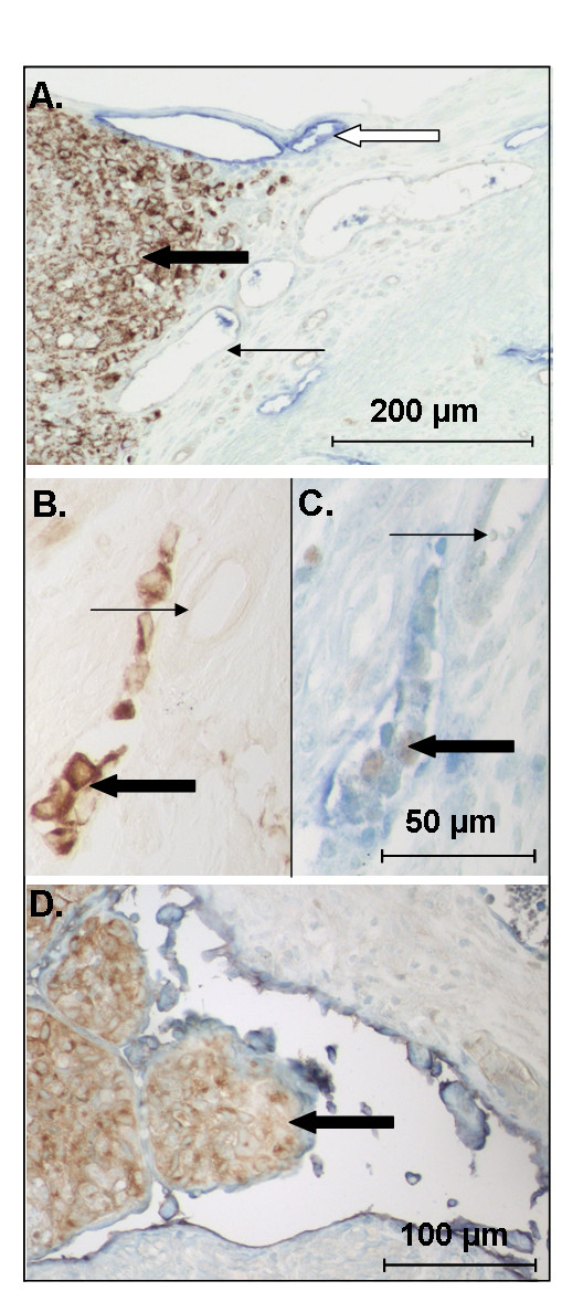
VEGF-D expressing 293EBNA cells invade uterine lymphatic vessels. Uterine horns of NOD SCID mice were inoculated with control or VEGF-D expressing 293EBNA cells. (A) Control tumor cells (brown) did not invade uterine blood or lymphatic (blue) vessels; however, (B-D) examples of myometrial lymphatic vessels containing tumor cells were observed in all mice treated with VEGF-D expressing cells. Note the tumor cells in a lymphatic vessel adjacent to unaffected blood vessels in B and C. Note the large tumor emboli in the distorted lymphatic vessel in D. (A and B: brown: tumor cells; C and D; blue: lymphatic vessels, brown: PCNA positive proliferating cells). Heavy black arrow: tumor cells. Fine black arrows: blood vessel. White arrow: lymphatic vessel.