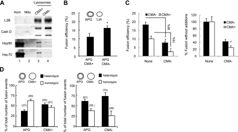
Figure 4.
Fusion events between APGs and different lysosomal subpopulations. A) Homogenate (Homo), mitochondria (Mito), and lysosomes with high (CMA+) and low (CMA−) CMA activity isolated from starved mice were subjected to immunoblots for the indicated antibodies. B) APGs CMA+ and CMA− lysosomes isolated from livers of starved mice were labeled with antibodies against LC3, hsc70, and LAMP-2B, respectively, and the corresponding fluorophore-conjugated secondary antibodies. Fractions were incubated for 30 min at 37°C, and fusion events were analyzed. Values are expressed as percentage of fusion events relative to the total number of particles in the field and are means + se of 3–5 different experiments. C) APGs and CMA+ and CMA− lysosomes labeled as in B were incubated alone or in the presence of unlabeled CMA− lysosomes, and fusion events were analyzed. Values are expressed as percentage of fusion events relative to the total number of particles in the field (left panel; percentage of decrease in the presence of unlabeled lysosomes is shown) or as percentage of the fusion efficiency in the absence of unlabeled CMA− lysosomes (right panel). Values are means + se of 3 different experiments. D) Homotypic and heterotypic fusion events in fusion reactions like those described in B for APGs with CMA+ lysosomes (left panel) or CMA− lysosomes (right panel). Values are expressed as percentage of total fusion reactions and are means + se of 3 different experiments. *P < 0.05.

