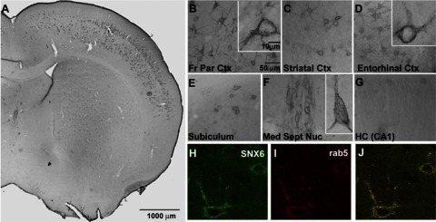
Figure 3.
SNX6 expression in mouse brain. A) Immunohistochemical analyses in brain tissue of C57Bl/6 mice show neuronal distribution in the cortical layers. B–G) Regional distribution of SNX6 in mouse brain: front parietal cortex (B), striatal cortex (C), entorhinal cortex (D), subiculum (E), medial septal nucleus (F), and hippocampus (G; CA). H–J) Subcellular colocalization with Rab5: SNX6 (H), Rab5 (I), and overlay (J). Note the predominant neuronal expression of SNX6 in indicated regions of the mouse brain.

