Fig. 2.
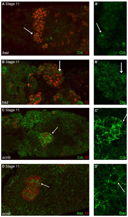
Tubule cell polarity is lost in both baz and scrib mutant embryos from the earliest stages of development. Confocal microscopy images of stage 11 baz maternal/zygotic (M/Z) (A,B) and scrib M/Z (C,D) embryos stained for Ct (red) to visualise the renal tubules. (A,B) In baz M/Z mutant tubules Crb levels are strongly reduced and any remaining protein is mis-localised (A,A′, arrow). Dlg has spread around tubule cell membranes (B,B′, arrow). (C,D) In scrib M/Z mutant embryos, both Crb (C,C′, green) and Baz (D,D′, green) are delocalised in tubule cells and can be found all around the cell cortex (arrows).

