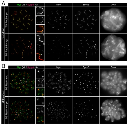
Fig. 5.
Normal chromosome development until metaphase I in TG Rec8-N-Myc spermatocytes. Immunofluorescence images of chromosome spreads stained with anti-Myc and anti-Sycp3 antibodies. (A) Pachytene nuclei and magnified sex chromosomes synapsed at the PAR (arrow) are shown. (B) Metaphase I chromosomes and magnified images of representative bivalents are shown.

