Fig. 6.
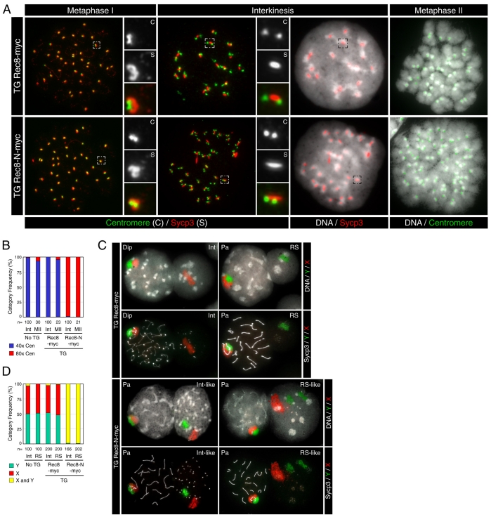
No nuclear division in spermatocytes expressing Rec8-N-Myc. (A) Immunofluorescence images of chromosome spreads stained with a CREST serum (for centromeres), anti-Sycp3 antibody and DAPI (for DNA). Small panels show the magnified images of boxed areas. (B) Frequencies of nuclei classified by number of centromeres: approximately 40 CREST foci (40× Cen) or approximately 80 CREST foci (80× Cen). Numbers of nuclei examined are indicated (n). (C) Chromosome paintings detecting X (red) and Y (green) chromosomes merged on DAPI images or on anti-Sycp3 images in grayscale. Stages are: Pa, pachytene; Dip, diplotene; Int, interkinesis; RS, round spermatid. (D) Frequencies of nuclei classified by distribution of sex chromosome painting signals: one patch of X chromosome alone (X), one patch of Y chromosome alone (Y) or one or two patches of both (X and Y). Numbers of nuclei examined are indicated (n).

