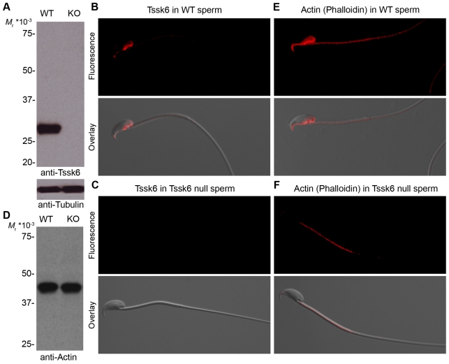
Fig. 5.
Tssk6 and F-actin localize to the posterior head and perforatorium of WT sperm. (A) Cauda epididymal sperm proteins from either WT or Tssk6-null (KO) mice were extracted, separated by polyacrylamide gel electrophoresis, transferred to PVDF and analyzed by western blot with anti-Tssk6 mAb. The signal was observed by incubation with horseradish-peroxidase-labeled anti-mouse IgG antibodies and chemiluminescence reaction. The same membranes were then probed with anti-tubulin antibodies as loading controls (lower panel). (B,C) Sperm from either WT (B) or Tssk6-null (C) mice were fixed and stained with anti-Tssk6 mAbs as described in the Materials and Methods. (D) Western blot reveals comparable levels of total actin of WT and KO sperm. (E,F) Epifluorescence localization of polymerized actin as stained with fluorescently labeled phalloidin in either WT (E) or knock-out (F) sperm incubated for 1 hour in media that support capacitation. In all cases, bottom panels show the overlay with a DIC image for orientation reference. Experiments were repeated at least three times; representative images are shown.

