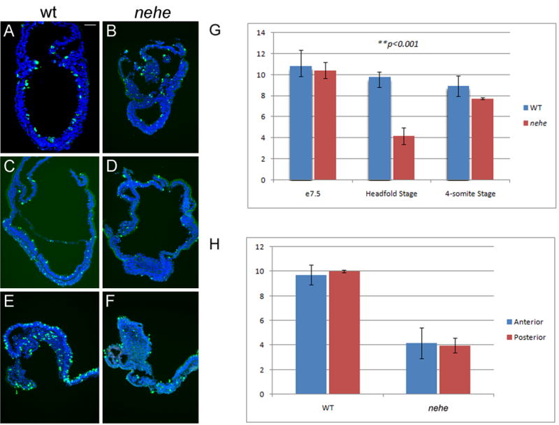
Figure 5. Reduced proliferation in headfold nehe embryos.

(A–F) Immunofluorescent staining with antibodies that recognize phospho-histone H3 (green) detects mitotic cells in wild-type (A, C, E) and nehe (B, D, F) embryos at e7.5 (A, B), headfold (C, D) and 4–6 somite (E, F) stages. Blue = DAPI staining. Anterior to the left. Scale bars: 100 μm in A–F. Note the strong reduction in the number of mitotic cells in the entire embryonic region of headfold-stage nehe embryos. (G) Phosphohistone H3-positive and total (DAPI-stained) nuclei were counted in sections from wild-type and mutant embryos at the three developmental stages. Embryonic and extraembryonic regions were counted separately. The only significant difference between wild type and mutant was in the embryonic region at the headfold stage, when the number of mitotic cells in the mutant was three-fold lower than in wild-type. These two values are different with p = 0.0001. At the same stage, there was no significant difference between the mitotic indices in the extraembryonic regions of wild type and mutant embryos. (H) The same sections from the headfold stage embryos analyzed in (G) were divided into two halves, anterior and posterior to the node, and phospho-histone H3 and DAPI-positive cells were counted. There was no significant difference in the mitotic index in the anterior versus posterior half of either wild-type or mutant embryos.
