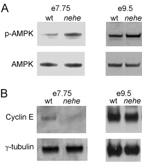
Figure 6. AMPK activation and decreased cyclin E in nehe embryos.

(A) Western blot analysis of phospho-AMPK in embryo extracts. Representative experiments are shown. Three independent samples for mutants and seven independent samples for wild type were analyzed; averaged among the experiments, the mutants had a 2.6+0.1-fold higher level of p-AMPK/total AMPK relative to wild type. At e9.5, three independent mutant protein extracts and seven independent wild-type extracts were analyzed, on average the p-AMPK/total AMPK ration in mutant embryos was 1.2±0.1-fold higher than in stage-matched controls. (B) Western blot analysis of cyclin E in headfold and e8.5 embryos; representative experiments are shown. Three independent mutant and five wild type samples were analyzed at headfold stage; the level of cyclin E in mutants (relative to the γtubulin loading control) was 0.38±0.04 the value in wild type. At e9.5, three mutant and five wild type extracts were analyzed; the level of cyclin E in mutants at this stage was equivalent to the amount in wild type (1.0±0.1). γtubulin was used as a loading control. Each lane in parts (A) and (B) represents protein extracted from 4–8 embryos.
