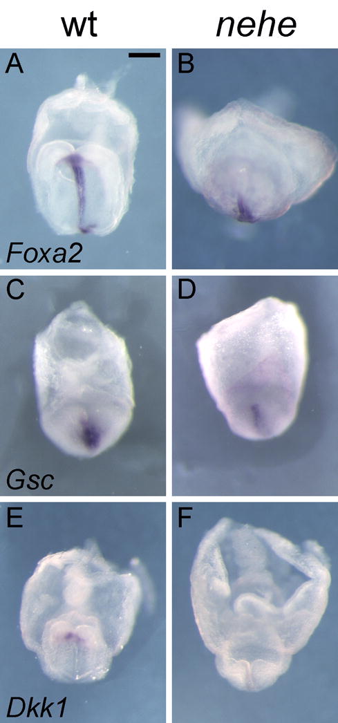
Figure 8. Loss of anterior neural fates and defective specification of the prechordal plate in nehe embryos.

(A–B) Foxa2 is expressed normally in the posterior midline but is barely detectable in the anterior midline of stage-matched nehe embryos (n=3), compared to stage-matched wild types. (C–F) The anterior mesendoderm markers Gsc (C, D) (n=6) and Dkk1 (E, F) (n=4) are barely detectable at the anterior mesendoderm of the mutant embryos, compared to stage-matched wild types. Scale bars: 0.25mm in A–H.
