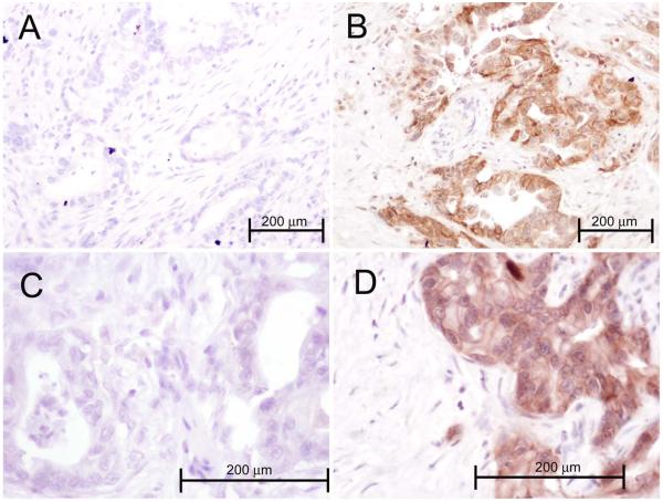
Figure 2.
The photographs show representative samples of pancreatic adenocarcinoma from 4 patients, stained using immunohistochemistry for EGFR. Each sections shows malignant glands composed of plump epithelial cells with nuclear enlargement and pleomorphism, and the intervening desmoplastic reaction of small fibroblasts. Panels A and C (magnification 200 and 400X, respectively) were scored as 0 (cytoplasmic), 0 (membranous). Panels B and D represent EGFR expressing cancers and were scored as 140 (cytoplasmic) and 60 (membranous). The stromal elements were negative in all samples.

