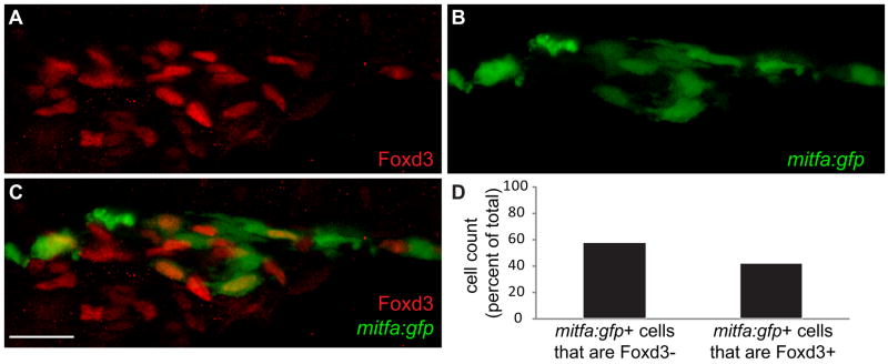
Fig. 4. mitfa positive neural crest cells re-acquire Foxd3 expression.
(A–C) Confocal images taken from lateral aspect of anterior trunk, 40X. (A) Foxd3, (B) mitfa:gfp, (C) Merge: Red channel: Foxd3. Green channel: mitfa:gfp. (D) Cell counts of mitfa:gfp positive cells that are either Foxd3 positive or negative, counts derived from 40X confocal images at 24 hpf, numbers given as percent of total. 52% of mitfa:gfp+ cells are Foxd3− (432/748). 48% of mitfa:gfp+ cells are Foxd3+ (316/748). Scale bar=30 μm.

