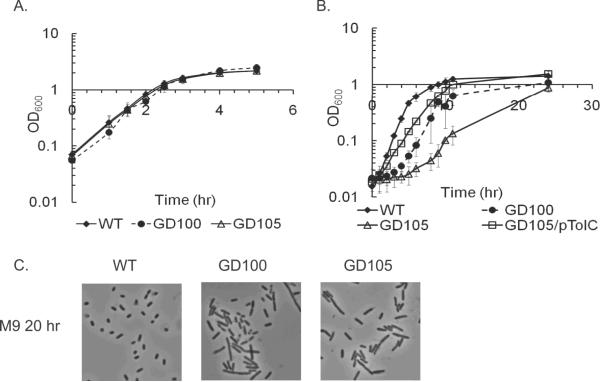
Figure 2.
Growth and cell division defects of ΔtolC cells. Overnight cultures of WT, GD100 (ΔtolC) and GD105 (ΔtolC–ygiBC ΔyjfMC) grown either in LB (A) or M9 (B) were re-inoculated (1:100) into the fresh matching medium. The growth was monitored by measuring absorbance at 600 nm. The initial cell densities were adjusted to approximately the same value. For each growth curve the average of three independent experiments is shown. Error bars are standard deviations (SD, n=3). C. Phase contrast microscopy. Cells were grown overnight in LB, diluted 1:100 into fresh M9 medium and incubated at 37°C for twenty hours.

