
Fig. 5.
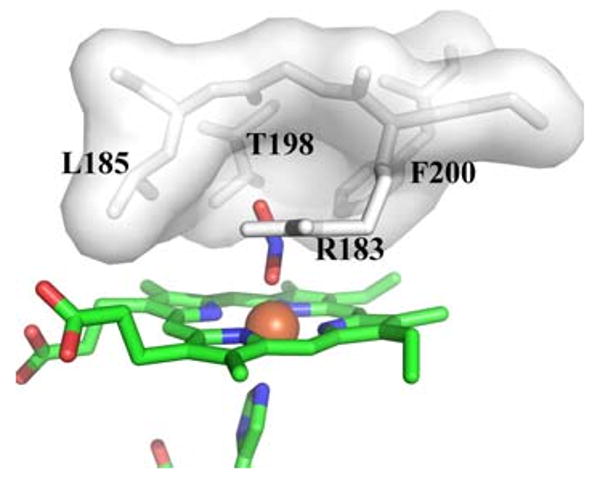
Distal pocket of Cld. Residues lining the distal pocket are shown as white sticks with a transparent surface, except for Arg-183. The heme and ligands are drawn as sticks colored by atom (carbon, green), with the iron as an orange sphere. This figure was generated using PyMOL (http://www.pymol.org/)