Figure 1.
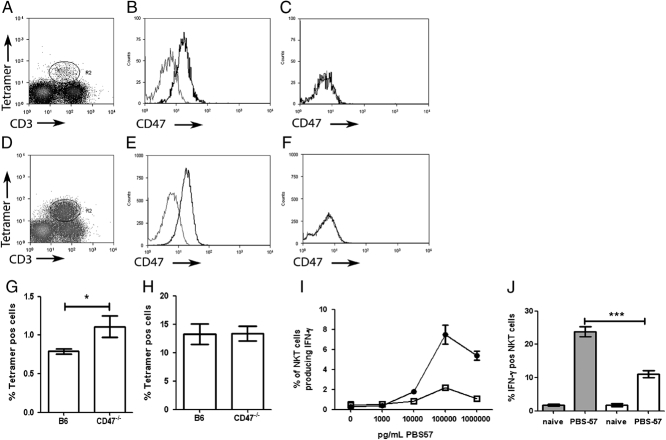
Phenotype, frequency and responsiveness of iNKT cells from CD47−/− mice. (A–F) Expression of CD47 on iNKT cells on spleen (A–C) and liver (D–F) iNKT cells. Similar profile of CD3 and tetramer staining were obtained for CD47−/− mice. Spleen (A) and liver (D) iNKT cells were identified by excluding autofluorescent cells and then gating on PBS-57-loaded CD1d tetramer+ CD3+cells. Splenic (B) and hepatic (E) iNKT cells in B6 mice express CD47, whereas splenic (C) and hepatic (F) iNKT cells in CD47−/− do not. Dotted lines represent isotype controls. (G–H) The percentage±SEM of tetramer+ cells in the spleen (G) and liver (H) of naïve B6 and CD47−/− mice. (n=20 individual mice from three independent experiments.) (I) IFN-γ production by splenic tetramer+ TCR-β+ cells after 16 h in vitro stimulation with PBS-57; C57BL/6 (closed circles) and CD47−/− mice (open squares). Data represent mean±SEM of triplicate samples pooled from three to five mice and are representative of three independent experiments. (J) IFN-γ production by splenic tetramer+ TCR-β+ iNKT cells 16 h after i.v. injection of 10 ng PBS-57. Data represent mean±SEM (n=8 mice from two independent experiments). *p<0.01, ***p<0.0001, Mann–Whitney U test.

