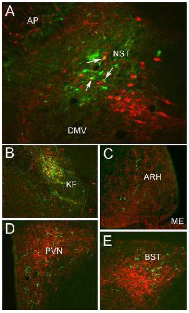
Figure 2.
Dual immunofluorescent localization of PhAL (green) and the noradrenergic synthetic enzyme, DbH (red). A: Individual NST neurons concentrating PhAL (green) within the iontophoretic tracer injection site (see Figure 1). A subset of these PhAL-positive neurons are DbH-positive (arrows point out 3 examples). B: PhAL-labeled fibers within the KF subregion of the lateral parabrachial nucleus. C: PhAL-labeled fibers within the hypothalamic ARH. D: PhAL-labeled fibers within the PVN. E: PhAL-labeled fibers within the BST. Note that each photomicrograph depicts PhAL and DbH immunofluorescent labeling photographed at only one focal plane through the section. See Table 1 for abbreviations.

