
Fig. 5.
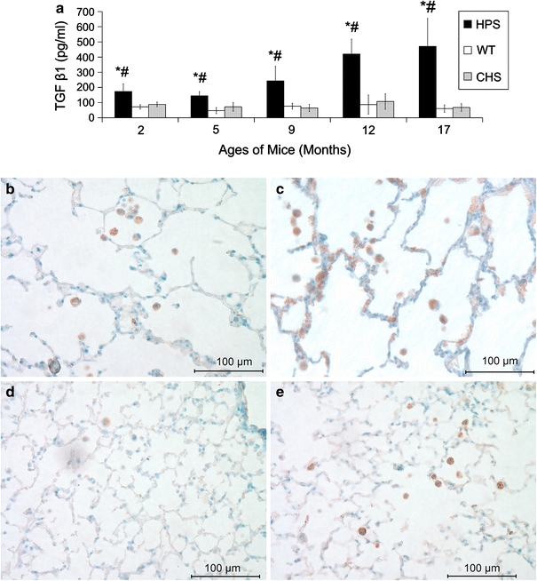
TGF β1 BAL levels and localization in lungs. Panel a shows average TGF β1 (±SD) levels in BAL samples of HPS, WT, and CHS mice from 2 to 17 months of age using ELISA. Samples of 4–10 mice were used for each time point and strain. Significant difference P < 0.05: *HPS versus WT; #HPS versus CHS. No significant difference between CHS and WT was found at any age tested. Panels b–e are representative TGF β1 IHC stained (brown precipitate) lung sections from HPS at (b) 9 and (c) 12 months of age and from (d) WT and (e) CHS at 12 months of age. AMs (intra-alveolar cells) were darkly stained in each of the mouse strains at all ages tested, and staining appeared in epithelial cells as well in older HPS mice but not in WT or CHS mice