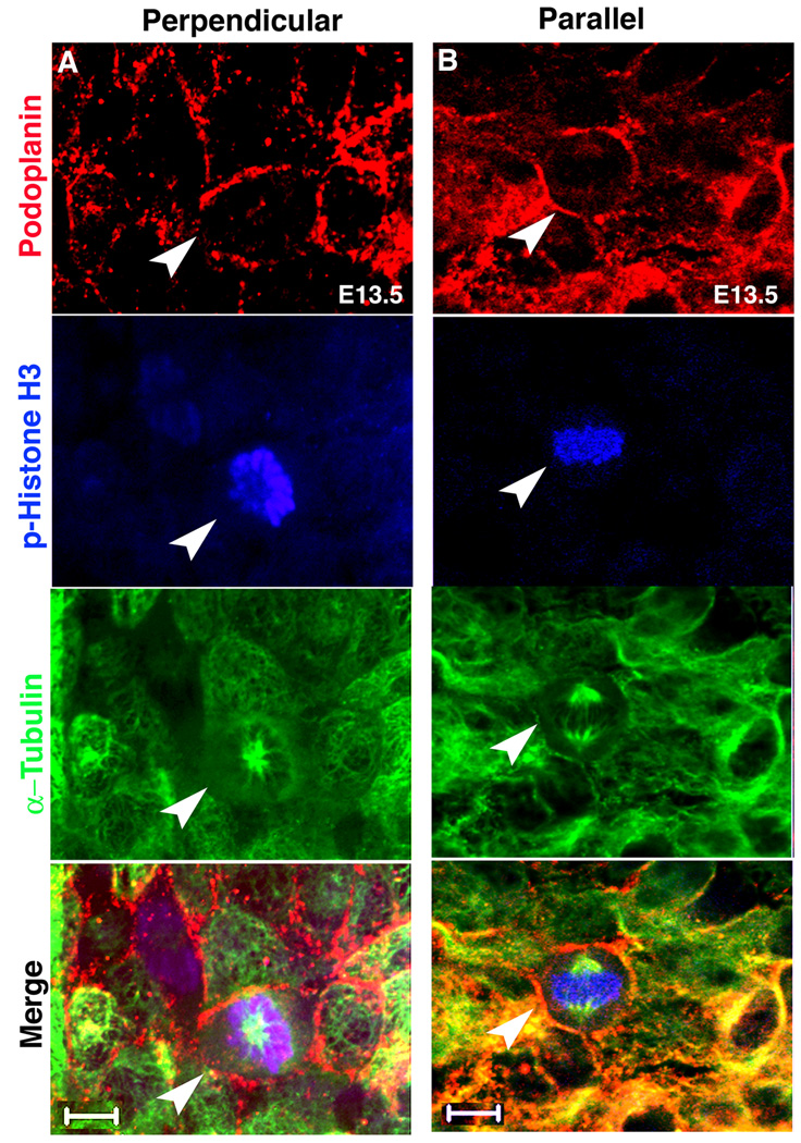
Figure 3. Whole mount staining of dividing epicardial cells.
(A) Perpendicular and (B) parallel divisions can be observed in whole mount stained hearts. Podoplanin, p-Histone H3 and α-Tubulin stained: epicardial cells, mitotic cells and spindles respectively. Arrowheads point to the mitotic cells. Scale bars = 10 µm. See Table S1 for additional information.

