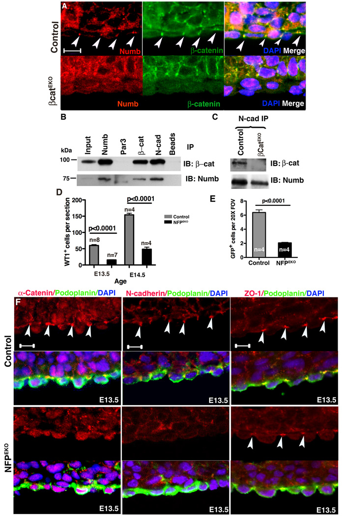
Figure 6. Numb function in adherens junctions and NFPEKO epicardial phenotype.
(A) Numb localized to adherens junctions in control but not in βcatEKO epicardial cells. Arrowheads indicate adherens junctions. (B) Numb, Par3, N-cadherin (N-cad) and β-catenin (β-cat) were immunoprecipitated from primary epicardial cell lysates and blotted for the indicated proteins. (C) N-cadherin was immunoprecipitated from control and βcatEKO epicardial cell lysates and blotted for interactions with the indicated proteins. Input was 5% of the lysate. (D) Fewer WT1+ cells were present in the myocardium of NFPEKO than in control hearts. (E) Ad- GFP was used to trace epicardial cell EMT in an ex vivo culture system. (F) Adherens junctions (α-catenin and N-cadherin) and tight junctions (ZO-1) in NFPEKO epicardial cells. Arrowheads point to junctions. Podoplanin was used to identify epicardial cells. Scale bar = 10 µm. See also figure S4.

