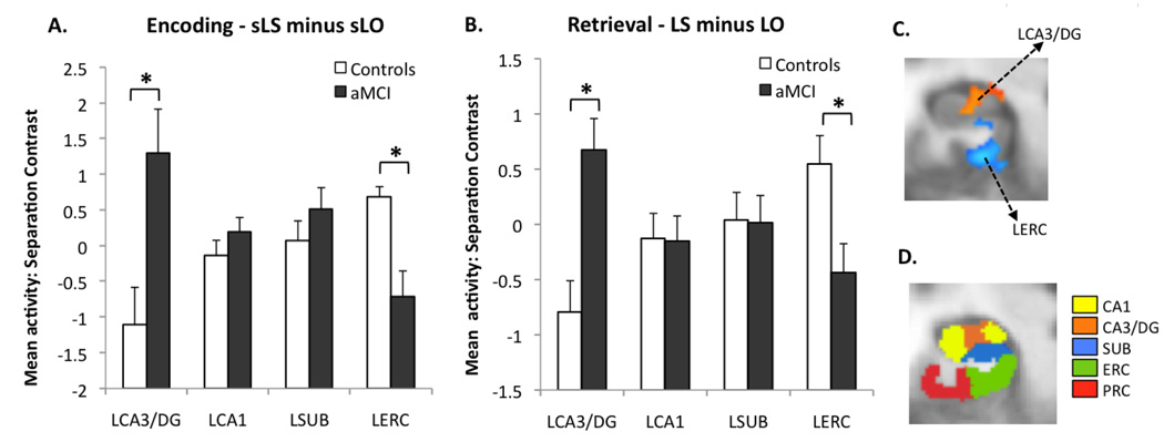
Figure 4.
Results of the critical contrasts. (A) During encoding (lures subsequently called “similar” minus lures subsequently called “old”) aMCI patients had significantly higher activity in the left CA3/DG and lower activity in the left entorhinal cortex. (B) During retrieval (lures called “similar” minus lures called “old”), there was an identical pattern of group difference. All bars are mean ± s.e.m. (C) The two ROIs of interest in this contrast during encoding shown on an average customized structural template based on the entire sample (functional ROIs based on the t-contrast between groups). Group difference in activity during retrieval was very similar. (D) A sample slice from the anatomical segmentation template in the left medial temporal lobe.

