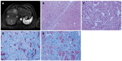
Figure 1.
Baseline radiologic and pathologic assessment of the patient in late 2006 before initiation of therapy. A: Initial magnetic resonance imaging (MRI) scan revealing multiple hepatocellular carcinoma (HCC) nodules; B: Regenerative nodule (lower right) juxtaposed to well circumscribed HCC (upper left) separated by a fibrous septum with a sparse lymphocytic infiltrate (magnification, × 100); C: Representative high power magnification of the HCC showed large tumor cells with occasional vacuoles and microvesicular fatty degeneration of hepatocytes (magnification, × 200); D: Abnormally increased microvessel density within the HCC as demonstrated by CD34 immunohistochemistry (magnification, × 200); E: Tumor cells express VEGFR1 at the tumor front (right) contrasting with weak expression towards to the tumor centre (left; magnification, × 200).

