
Figure 3.
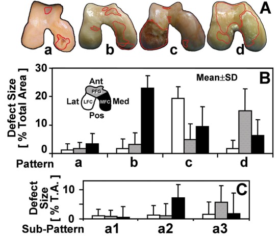
(A) representative photographs of the samples within 4 main patterns: a, mixed; b, medial femoral condyle; c, lateral femoral condyle; d, patellofemoral groove. Location-dependent size of full-thickness lesions of samples classified within (B) 4 main patterns and (C) subpatterns of a: a1, mixed small; a2, medial femoral condyle small; a3, patellofemoral groove small. n = 12 to 68 (see Table 1).