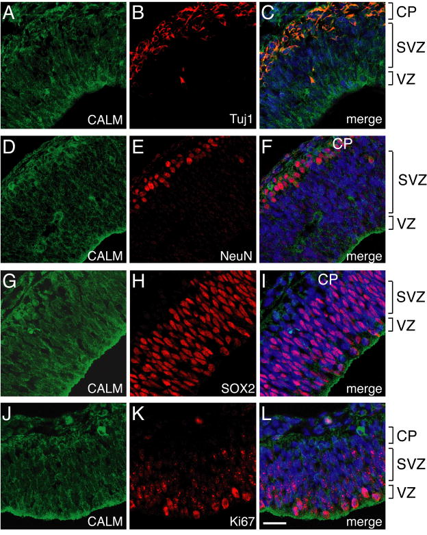
Figure 4. CALM is present in differentiated neurons and also in neural progenitors.
A-F: Double staining of CALM (antibody sc6433) with Tuj1 (A-C) and NeuN (D-F) showing CALM’s presence in young and mature neurons. G-L: CALM was also present in SOX2-positive neural progenitors (G-I) and Ki67-positive proliferating cells (J-L). Scale bar: 20 μm (applies all images). Results of CALM staining using a different CALM antibody (antibody sc5395) are shown in Supplementary Figure S4. CALM’s diffuse staining pattern did not permit quantitative analysis as used in Figure 2.

