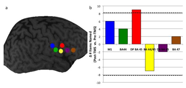
Figure 1.

a) The six sites at which TMS was delivered in Phase 1 of the study: M1 corresponding to the mouth (blue), Brodmann area 44 (BA 44; green), dorsal posterior BA 45 (red), dorsal anterior BA 44/ventral posterior BA 45 (yellow), dorsal anterior BA 45 (purple), and BA 47 (brown).
Figure 1b) The difference in performance on a naming task before and after rTMS to the six right hemisphere sites. Hatched lines indicate significant differences.
