
Figure 3.
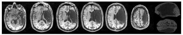
T1-weighted MRI (1mm slice images with field of view 192 × 256 × 80; axial images depicted are—from inferior to superior—slices 124, 100, 90, 84, 78, and 66). Extensive cortical lesion is present in middle and inferior frontal gyrus areas (includes Broca’s area), and in most of the middle and superior temporal gyrus areas (includes Wernicke’s area). There is some sparing of deep white matter, adjacent to inferior frontal horn (arrows, medial subcallosal fasciculus), deep to Broca’s area. Extensive sensorimotor cortex and parietal lobe lesion is present, with sparing of parts of BA 37 and 39 (angular gyrus).