Figure 3.
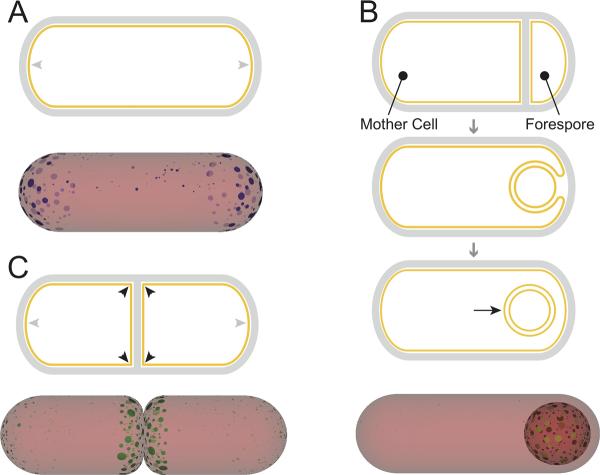
Curvature sensing of geometric differences in a rod-shaped bacterium. (A) Polar localization: above, a rod shaped cell is depicted in which the hemispherical cell poles (grey arrowheads) are the preferred localization sites for the lipid cardiolipin. Below, a schematic of results obtained from a simulation in which molecules with a preference for curved surfaces localize via cooperative interactions. By forming clusters, molecules with a preference for negative curvature (shown in blue, representing cardiolipin) can distinguish cell poles (grey arrowheads) from the cylindrical region of the cell membrane (red). (B) Stages of sporulation in B. subtilis: in the top panel, a cell divides asymmetrically to produce the larger mother cell and the smaller forespore. In the middle panel, the mother cell begins to engulf the forespore, which causes the septum to curve. Finally, in the bottom panel, the forespore pinches off as a double-membrane bound organelle, the cytosolic face of which is the only positively curved surface (black arrow) in the mother cell cytosol. Below, by forming clusters, molecules with a preference for positive curvature (shown in green, representing SpoVM) might be able to distinguish the outer spore surface from the rest of the cytosolic surface. (C) Hierarchical curvature localization: above, depiction of an actively dividing B. subtilis cell in which sharp membrane inflections created by a nascent division septum (black arrowheads) are the primary site of DivIVA localization, whereas the hemispherical cell poles (grey arrowheads) are the secondary site of DivIVA localization. Below, by forming clusters, molecules with a preference for negatively curved surfaces (representing DivIVA) might be able to distinguish between the highly curved division septum (black arrowheads), the less curved hemispherical poles (grey arrowheads), and the least curved lateral edge of the cell.

